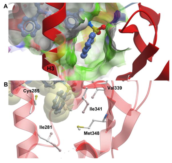
Figure 6.
SR2067 stabilization of the β-sheet region. The PPARγ LBD is shown as ribbons (red) and SR2067 is represented as sticks with carbon atoms colored blue. (A) Surface representation of the β-sheet pocket with green color used to signify hydrophobicity. (B) Hydrophobic contacts of SR2067 with the PPARγ β-sheet region. SR2067 is shown in sticks and with transparent van der Waals radii. PPARγ residues engaged in hydrophobic contacts have side chains displayed as sticks.

