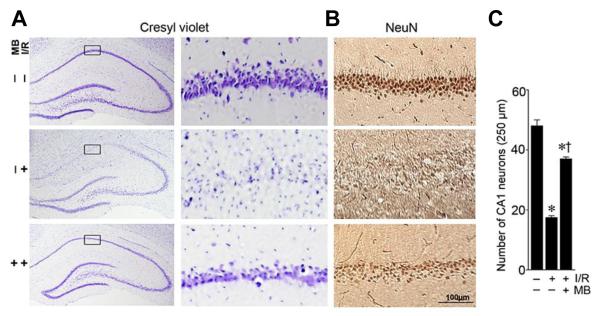
Fig.1.
Treatment with MB results in significant neuroprotection in hippocampal CA1 following GCI. A: Cresyl violet staining shows the whole hippocampal overview and the detailed CA1 region. B: High magnification DAB staining with anti-NeuN antibody shows pyramidal neurons in medial hippocampus CA1 region (scale bar: 100μm). C: The number of surviving neurons per 250-μm length of medial CA1 were quantitated and statistically compared. Data are presented as mean ± SE, n= 11 per group. * P<0.05 vs. sham, † P<0.05 vs. ischemic reperfusion (I/R) group.

