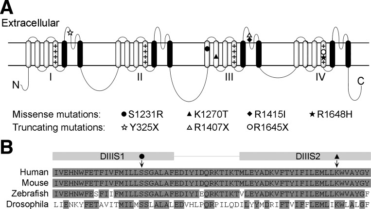
Fig. 1.
A: schematic of the structure of a voltage-gated sodium channel α-subunit. Each of the 4 domains (I–IV) contains 6 transmembrane segments (S1–S6, from left to right). Plus sign (+) columns indicate voltage-sensing transmembrane segment S4 in each domain; S5 and S6 form the channel pore (black). The locations of the 7 SCN1A mutations that are referred to in this review are denoted by symbols. B: aligned amino acid sequence from the transmembrane segment S1 to S2 in domain III (DIII) of human, mouse, zebrafish, and fruit fly (Drosophila). The locations of the S1231R (solid circle) and K1270T (solid triangle) mutations are indicated by arrows.

