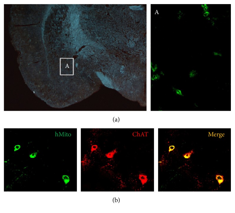
Figure 4.

Representative images for detection of transplanted F3.ChAT cells in the amygdala (a) and for production of functional ChAT protein (b) 4 weeks after transplantation. ((a), inset A) For detection of human F3.ChAT cells, the amygdala tissue was immunostained with hMito antibody. (b) The cells were double immunostained with antibodies specific for hMito and ChAT. NMDA, N-methyl-d-aspartate; hMito, human mitochondria; ChAT, choline acetyltransferase.
