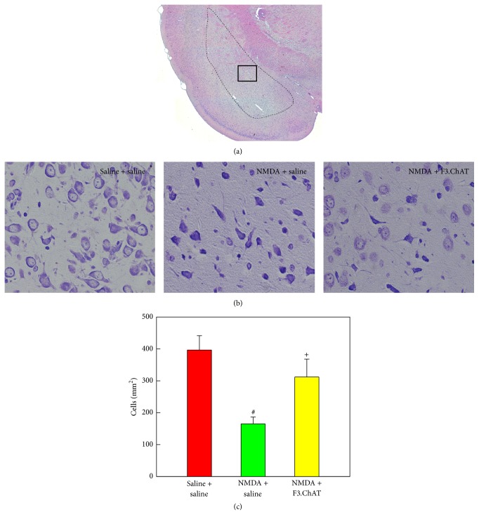
Figure 5.
Representative hematoxylin-eosin (a) and Nissl staining (b) images of the amygdala, and the cell number in the amygdala (c) 4 weeks after transplantation. After examination on the entire amygdala (dotted line in (a)), magnified medial amygdala (inset in (a)) was photographed (b), and the number of survived neurons was counted (c). NMDA, N-methyl-d-aspartate. #Significantly different from vehicle control (P < 0.05). +Significantly different from NMDA alone (P < 0.05).

