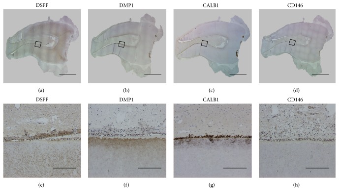
Figure 4.
Immunohistochemical (IHC) staining of dental pulp tissue. IHC staining for DSPP (a, e). DSPP was noted in pulpal tissue, the odontoblast layer, and primary and secondary dentin. IHC staining for DMP1 (b, f) and CALB1 (c, g). CALB1 was especially expressed in pulpal tissue and the odontoblast layer. IHC staining for CD146 (d, h) (scale bars: (a)–(d) 4 mm, (e)–(h) 200 μm).

