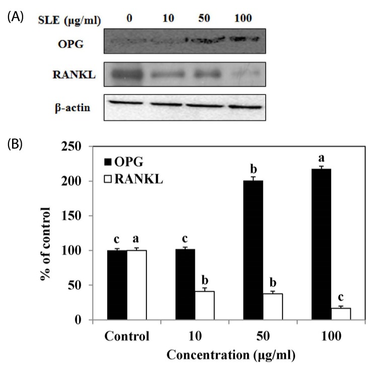
Fig. 5. Effects of SLE on the OPG/RANKL ratio in MC3T3-E1 cells.
Cells were treated with SLE at 10, 50 and 100 µg/ml for 2 days. (A) The protein expression of OPG/RANKL ratio, such as OPG and RANKL was detected by western blot. (B) Relative expression was quantified by densitometry using the Multi Gauge V3.1 and calculated according to the reference bands of β-actin. Each value is expressed as mean ± SD (n = 3). a-cValues with different letters were significantly different at P < 0.05, as analyzed by Duncan's multiple range test. SLE: Scytosiphon lomentaria extract.

