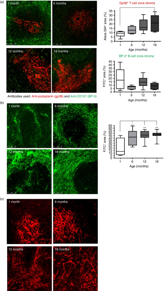
Figure 2.

Stromal changes within spleens of young and old mice. (a) Sections were stained with the anti‐gp38 (podoplanin; in red), which detects the T‐cell stroma, and anti‐CD157 (BP‐3), which detects the B‐cell stroma, (shown in green). The level of staining was assessed by imageJ analysis (graphs on the right hand side) and revealed an increase in the T‐cell stromal area. Magnification ×100. (b) Staining for the fibroblastic reticular cell (FRC) with ERTR7 antibody revealed an increased deposition of FDC around the marginal zone in the splenic sections from older mice. Magnification ×100. (c) Staining for follicular dendritic cells (FDC) using anti‐FDC‐M2 revealed that the network of FDC appears more dispersed in the splenic sections from older mice Magnification ×200. Isotype controls revealed no staining (data not shown). Data representative of four experiments. *P < 0·05; **P < 0·01.
