Figure 1.

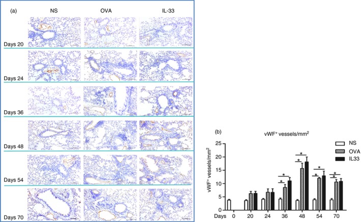
Interleukin‐33 (IL‐33) increased airways von Willebrand factor (vWF) immunoreactivity. (a) Representative photomicrographs of vWF immunoreactive blood vessels in lung sections from saline‐ (NS), ovalbumin‐ (OVA) and IL‐33‐challenged mice at various time‐points as indicated (light microscopy, × 200). (b) Quantitative analysis of numbers of vWF + blood vessels per unit area of lung sections. Data are expressed as mean ± SEM (n = 5 mice in each group at each time‐point). *P < 0·05.
