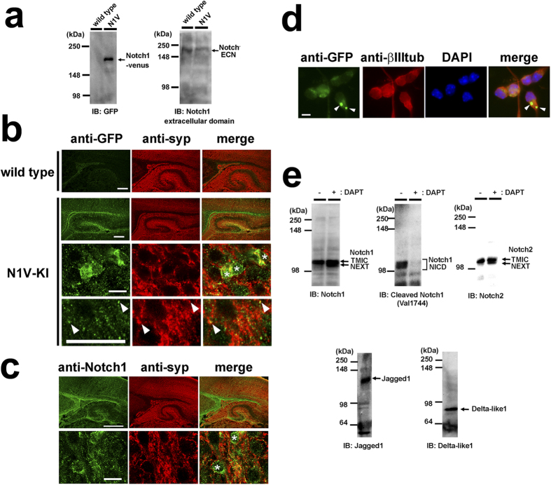
Figure 1. The expression of Notch and Notch ligands in mouse brain and primary neuronal culture.
(a) Immunoblot analysis of Notch1-Venus protein in N1V-KI mouse brain. Brains of wild type or homozygous N1V-KI mice at postnatal day (P) 1 were homogenized and analyzed by immunoblotting using antibodies against GFP (left, TMIC of Notch1-venus) and Notch1 extracellular domain (right, ECN). (b) Representative images of sagittal sections of cerebral cortex from P1 wild type mouse or P0 homozygous N1V-KI mouse immunostained with anti-GFP (green) and anti-synaptophysin 1 (syp, red) antibodies. The bottom two panels of N1V-KI show a magnified image of cerebral cortical region. Asterisks indicate Notch1-positive cell bodies. Arrowheads indicate N1V-positive puncta colocalized with synaptophysin 1. Scale bars, 200 μm (the top two panels) and 10 μm (the bottom two panels). (c) Representative images of sagittal sections of cerebral cortex from wild type mouse at P0 immunostained for Notch1 intracellular domain (green) and synaptophysin 1 (red). Scale bar, 200 μm. The bottom panels are a magnified image of cerebral cortex. Scale bar, 10 μm. Asterisks indicate Notch1-positive cell bodies. (d) Expression of Notch1-Venus protein in 2 DIV cultured cortical neurons obtained from homozygous N1V-KI mouse immunostained with antibodies against GFP (green), neuron-specific β-III tubulin (red). Nuclei are co-stained with DAPI (blue). Notch1-Venus-positive puncta in soma are shown by arrowheads. Scale bar, 10 μm. (e) Immunoblot (IB) analyses of Notch1, Notch2, Jagged1 and Delta-like1/4 in 6 DIV primary neuronal culture obtained from wild type brains. Note that treatment with the γ-secretase inhibitor DAPT caused accumulation of NEXT of Notch1 and Notch2 and diminished the Notch1 NICD production.

