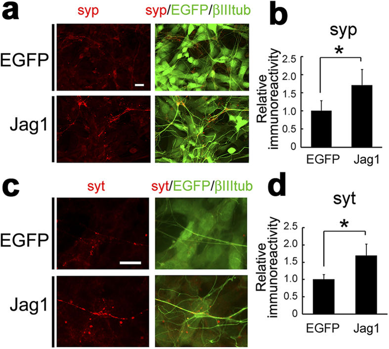
Figure 3. Induction of functional presynaptic terminals by Jagged1.
(a) Primary cortical neurons cocultured with EGFP-3T3 or Jag1-3T3 were immunostained with antibodies against synaptophysin 1 (red) and β-III-tubulin (green). Note that Jag1-3T3 cells also express EGFP by IRES-mediated translation (green). (b) Quantification of immunofluorescence signals of synaptophysin 1 (n = 4, means ± SEM. *p < 0.05 by student’s t-test). (c) Immunostaining of internalized antibody against synaptotagmin 1 (red) after depolarization in cocultured primary neurons. (d) Quantification of immunofluorescence signals of synaptotagmin 1 (n = 5, means ± SEM. *p < 0.05 by student’s t-test). Scale bars, 10 μm.

