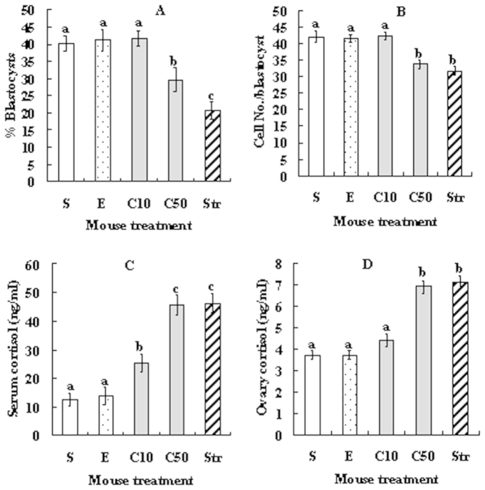
Figure 1. Effects of cortisol injection on blastocyst development of Sr2+-activated oocytes and on cortisol levels in serum and ovarian homogenates.
(A) % Blastocysts; (B) Cell number per blastocyst; (C) Cortisol in serum; and (D) Cortisol in ovary. At 24 h after eCG injection, experimental mice were injected with cortisol at 10 (C10) or 50 (C50) mg/kg body weight, control mice were injected with either saline (S) or ethanol (E), and stressed control (Str) mice were restrained for 24 h. For oocyte maturation, each treatment was repeated 5 times with each replicate containing about 30–35 oocytes from 2 mice. For cortisol assays, each treatment was repeated 3 times with each replicate containing ovarian homogenates or serum from 3 mice. a–c: Values without a common letter above bars differ significantly (P < 0.05).

