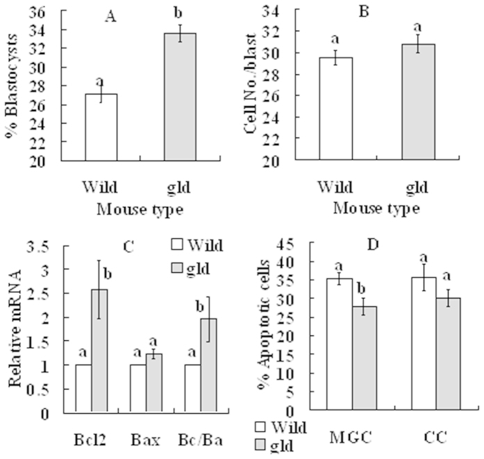
Figure 6. Effects of cortisol injection on blastocyst development and apoptosis in MGCs and CCs in wild-type mice and gld mice.
(A) Rates of blastocysts; (B) Cell number per blastocyst; (C) Levels of Bcl2 and Bax mRNAs and Bcl2/Bax (Bc/Ba) ratio in MGCs; and D. Percentage of apoptotic MGCs and CCs after Hoechst staining. At 24 h after eCG injection, mice were injected with 50 mg/kg cortisol, and at 24 h after cortisol injection, mice were sacrificed to collect ovaries for further experiments. Percentages of blastocysts and cell number per blastocyst were observed after Sr2+-activation of oocytes. Apoptotic percentages of CCs were observed after oocytes were cultured for 14 h without SGH. For oocyte maturation for embryo development, each treatment was repeated 3 times with each replicate containing about 30–35 oocytes from 2 mice. In experiments with MGCs, each treatment was repeated 4 times with each replicate including MGCs from 3 mice. For Hoechst staining of CCs, each treatment was repeated 3 times with each replicate containing CCs from 30 oocytes from 3 mice. a,b: Values without a common letter above their bars differ significantly (P < 0.05).

