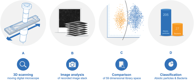
Figure 1. The various steps in determining the concentration of bacteria and abiotic particles.
(A) Schematic of flow cell, light source, lens, and camera. (B) Image stack of a particle coming into focus and out again as the tilted image plane moves across it. (C) Extraction of parameters from recorded image stacks and comparison to library data. (D) Classification of particles in “Bacteria” and “Abiotic particles”.

