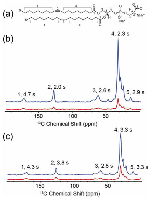
FIGURE 4.
1H-13C Cross Polarization 1D 13C NMR spectra of GALN and GCDL in protonated lipids. (a) 1,2-Dioleoyl-sn-Glycero-3-Phosphoethanolamine (DOPE), the dominant lipid in the vesicles. Numerical labels correspond to resonances identified in spectra of (b) GALN and (c) GCDL prepared with 1:3.5 protein:lipid ratio in vesicles of 9:1 DOPE:DOPS. Blue traces are DNP-enhanced spectra upon microwave irradiation while red traces are the same experiment without microwave irradiation. Spectra appear similar because they are dominated by lipid signals. In the case of GALN (b) the patterns of T1 obtained from a 3D inversion recovery array, peaks 2, 4, and 5 exhibit shorter T1 relaxation times compared to peaks 1 and 3, suggesting the nitroxide spin labels are situated in the middle of the bilayer, as predicted by the IMAG crystal structure. In the case of GCDL, the opposite trend is observed. All spectra were processed with 30 Hz of Gaussian line broadening. t

