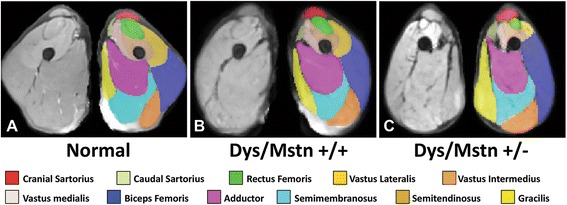
Fig. 5.

Averaged MRI segmentation of dogs from the three groups. Averaged T2-FS MRI images of pelvic limb muscles in the transverse plane at the level of the midthigh are shown in non-dystrophic control (a) dystrophic GRMD Mstn +/+ (b) and GRippet Mstn +/− (c) dogs. Note the proportional enlargement of the sartorius and hamstring muscles and the associated atrophy/hypoplasia of the quadriceps femoris of the dystrophic GRMD Mstn +/+ dogs, relative to the non-dystrophic control dogs, and the even more dramatic differential size of these muscles in the GRippet Mstn +/− dogs (also see quantitative measurements in Additional file 2: Table S1)
