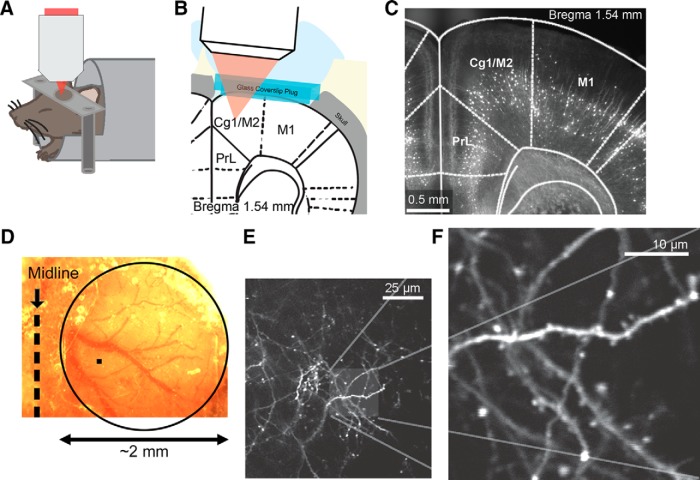
Figure 1.
Longitudinal imaging of dendritic architecture in the mouse medial frontal cortex. A, Schematic of the imaging experiment. B, Schematic of the long-term window implant. C, Fluorescence image of a fixed coronal brain slice from a Thy1-GFP-M mouse following longitudinal imaging. Cg1 and M2 (i.e., the MFC) were imaged in this study. PrL, prelimbic cortex. M1, primary motor cortex. D, Bright-field image of the long-term window implant. The glass window has an ∼2-mm-diameter width (circle), which is much larger than the imaging field of view of ∼60 × 60 μm (filled square). E, A low-magnification, in vivo two-photon image from layer 1 of the MFC in a Thy1-GFP-M mouse. Distal apical tuft branches from GFP-expressing layer 5 pyramidal neurons were visible. F, A high-magnification image of a region in E.

