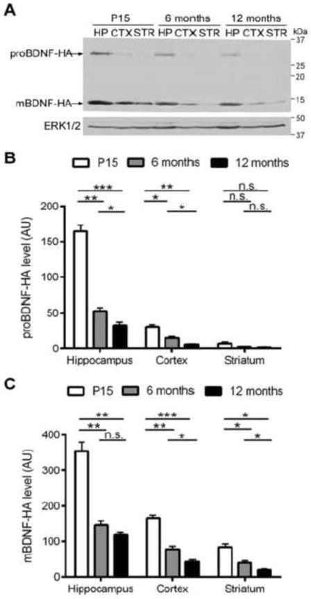
Figure 1. mBDNF is the predominant form and its level declines with age.
(A) Representative Western blot showing levels of BDNF isoforms in the hippocampus (HP), the cortex (CTX), and the striatum (STR) of wild type bdnf-HA/bdnf-HA mice at the age of postnatal day 15 (P15), 6 months and 12 months. Note that levels of both BDNF isoforms decrease with age, and intact proBDNF is barely detectable in the striatum at all the time points. Quantitative results of mBDNF level (B) and proBDNF (C) in HP, CTX and STR at the age of P15, 6months and 12 months show that the levels of both BDNF isoforms in CTX and STR are substantially lower than the HP, and both proBDNF and mBDNF levels decline significantly with age in all the three brain regions, except that proBDNF cannot be detected in STR at all the time points. AU, arbitrary units.

