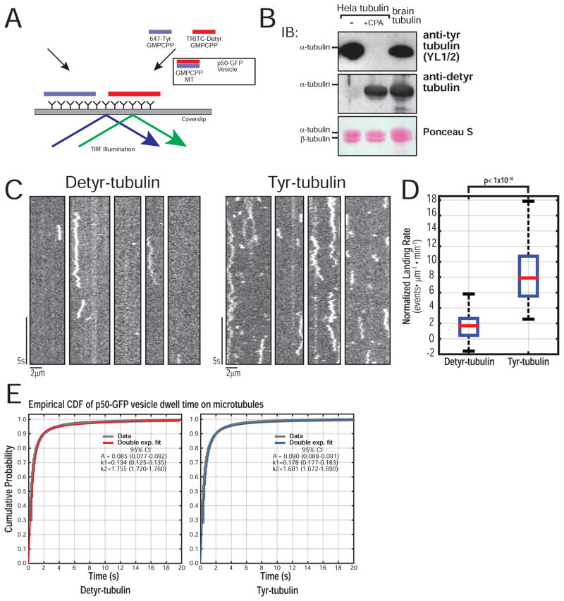
Figure 5.
Tyrosinated α-tubulin promotes robust organelle recruitment in vitro. (A) Schematic of the p50-GFP-vesicle recruitment assay using GMPCPP stabilized Tyr-/ Detyr-microtubules labeled separately with either 5% AF-647 or TRITC. (B) Western of Tyr/ Detyr tubulin purified from HeLa cells shows a homogenous population of fully tyrosinated or fully detyrosinated tubulin, compared to brain tubulin. (C) Representative kymographs of p50-GFP-vesicle landing on Detyr versus Tyr microtubules. (D) Quantification of landing rate shows a robust and significant increase in landing rate on Tyr microtubules (n = 70 movies per condition, each movie with > 500 events; N = 3 independent vesicle isolations; Wilcoxon Rank-Sum test p < 1×10⁻¹⁰). (e) Empirical CDF for microtubule dwell times of all non-fixed particles with double exponential fit and 95% CI for fit parameters. The dwell time distributions for vesicles on Tyr or Detyr microtubules were compared using a two sample KS test (p = 0.07; n > 1×10⁴ particles per condition, N = 3).

