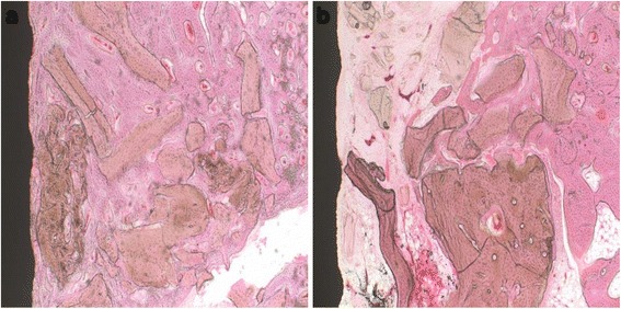
Fig. 5.

Histological sections at the sixth week. a Control group: defects treated with xenograft alone. b Experimental group: defects treated with xenograft and PRP. In the control group, the newly formed bone completely filled the bone gap; while in the experimental group, the newly formed bone filled 2/3 of the bone gap. Original magnification ×100. H&E stain
