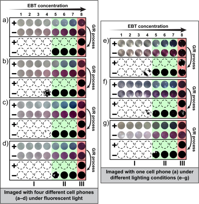
Figure 3.
Validation of the robustness of the G/R ratiometric approach to different hardware (cell phone cameras) and lighting conditions. (a–g) Enlarged and cropped color images (top two rows of each individual panel) captured by an unmodified cell phone camera from positive (+) and negative (−) RT-LAMP reactions at 2-fold increases in EBT concentration from 10.9 μM to 1.4 mM (1 = 0.011 mM; 2 = 0.022 mM; 3 = 0.044 mM, 4 = 0.088 mM, 5 = 0.175 mM; 6 = 0.35 mM; 7 = 0.7 mM; 8 = 1.4 mM). Positive wells are blue and negative wells are purple. After G/R ratiometric processing (bottom two rows of each individual panel), negative wells are black. Regions I, II, III in each panel indicate the effect of dye concentration: (II) acceptable concentration range for visualization (green regions); (I) concentrations too low for visualization (white regions); and (III) concentrations too high for visualization (red regions). (a–d) Images captured by four common cell phones under fluorescent light: (a) Apple iPhone 4S, (b) HTC inspire 4G, (c) Motorola Moto G, and (d) Nokia 808 PureView. (e–g) Images captured by an Apple iPhone 4S under three additional light conditions: (e) incandescent light, (f) direct sunlight, and (g) indirect sunlight. All experiments were performed with HCV RNA as a clinically relevant target. All images were acquired with unmodified cell phone cameras. Detailed information for the G/R ratiometric process (Figure S2) and additional cell phone camera images (Figure S3) are provided in the Supporting Information.

