Fig. 2.
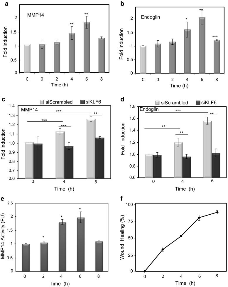
MMP14 protein expression and activity are increased during wound healing. MMP14 (a) and endoglin (b) protein levels on the surface of HUVECs at different time points during wound healing experiments were analyzed by flow cytometry as described in “Materials and methods.” Histograms indicate protein levels represented as fold induction with respect to resting cells at 8 h (control, C). MMP14 (c) and endoglin (d) protein levels on the surface of HUVECs at different time points after wound healing were measured by flow cytometry upon KLF6 suppression with siRNA specific for KLF6 (siKLF6). Scrambled siRNA (siScrambled) was used as a control. Histograms indicate protein levels represented as fold induction with respect to cells at 0 h. e MMP14 activity was measured during endothelial wound healing using a fluorogenic peptide and represented as a fold induction with respect to cells at time 0 h. f The percentage of endothelial healing is represented at different time points after wounding. Cells at approximately 80 % confluency (6 h after wounding) showed the highest MMP14 activity. *p < 0.05; **p < 0.01; ***p < 0.001 with respect to control condition (a, b), to time 0 h (e), or as indicated (c, d)

