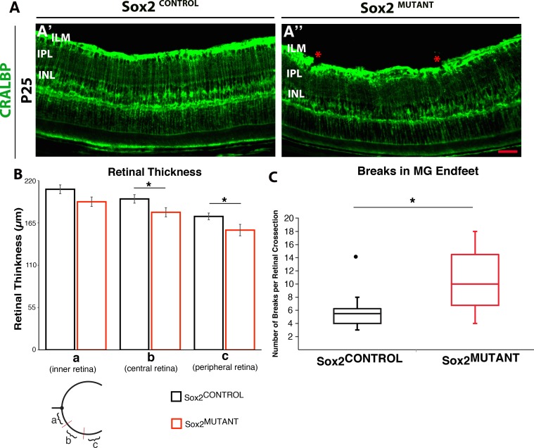
Figure 3.
Sox2 ablation reduces retinal thickness and results in disruption of the MG endfeet at the ILM at P25. (A) Staining of CRALBP demonstrates MG processes in the IPL are disorganized and their endfeet at the ILM are disrupted in Sox2MUTANT compared with Sox2CONTROL retinas. This phenotype was quantified in retinas stained with MG-specific GS. (B) The central and peripheral retina is significantly reduced in thickness in the Sox2MUTANT compared to Sox2CONTROL (average retinal thicknesses of the central retina 179 μm [SD ± 12] and 196 μm [SD ± 11], respectively, and the average retinal thicknesses of the peripheral retina is 156 μm [SD ± 15] and 173 μm [SD ± 9], respectively n = 5). *P values are: inner, 0.06; central, 0.04; peripheral, 0.05. (C) Sox2CONTROL retinas had significantly fewer disruptions of the MG endfeet compared to Sox2MUTANT retinas stained with GS (Average number of disruptions is 6 [SD ± 1.5] and 11 [SD ± 10.8], respectively n = 5). *P = 0.05. The • symbol in Sox2CONTROL represents a single outlying value not included in the statistical comparison. If the outlier is included, P = 0.07.

