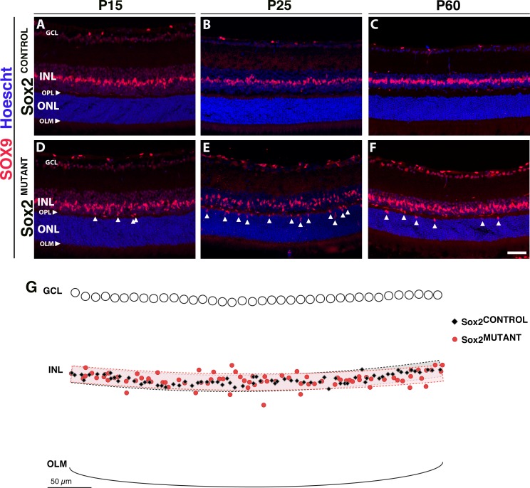
Figure 5.
Sox2MUTANT–Müller glial cell bodies are disorganized. (A–F) Müller glial cell bodies in the INL are specifically labeled with SOX9. (A, D) At P15, Sox2MUTANT MG (D) have mislocalized cell bodies compared with Sox2CONTROL (A), with some nuclei located ectopically in the OPL and the ONL (D, arrows). (B, E) At P25, Sox2MUTANT–MG cell bodies (E, arrows) display increased disorganization compared with Sox2CONTROL (B). Significant INL disorganization of Sox2MUTANT–MG cell bodies was identified by a least squares regression analysis (P = 0.042, n = 5 eyes). (G) Exported and overlaid (x, y) coordinates of a representative P25 Sox2MUTANT and Sox2CONTROL pair with graphical representation of the organizational bands used in the analysis. (C, F) At P60, Sox2MUTANT SOX9-positive MG remain disorganized (F, arrows) compared with Sox2CONTROL (C), and are decreased in number compared to the P25 Sox2MUTANT (E). Scale bar: 50 μm. Hoescht dye was used to stain the nuclei (blue).

