
Figure 1.
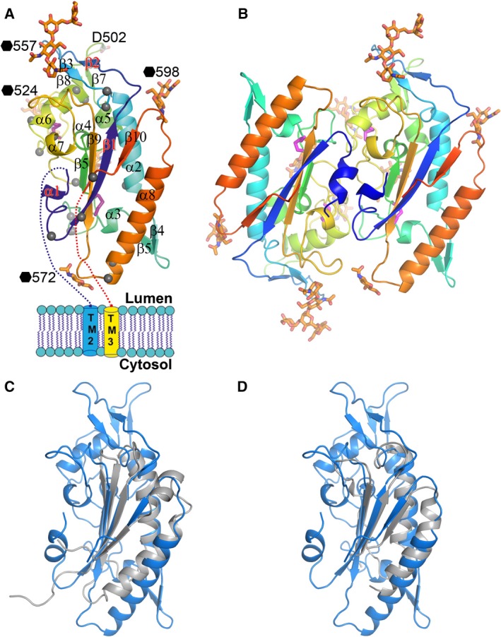
Overall structure of NPC1DC. (A) Cartoon representation of NPC1DC rainbow coloured from N (blue) to C‐termini (red). Grey spheres indicate disease mutation sites. Glycans are shown as orange sticks (labelled with black haxagons), disulphides as purple sticks. (B) The dimer in the crystallographic asymmetric unit. (C) Superimpositions of NPC1DC (blue) with the pore domain of bacterial multidrug efflux transporter MexB (grey) and (D) with the domain 2 of MmpL11 (grey).