
Figure 4.
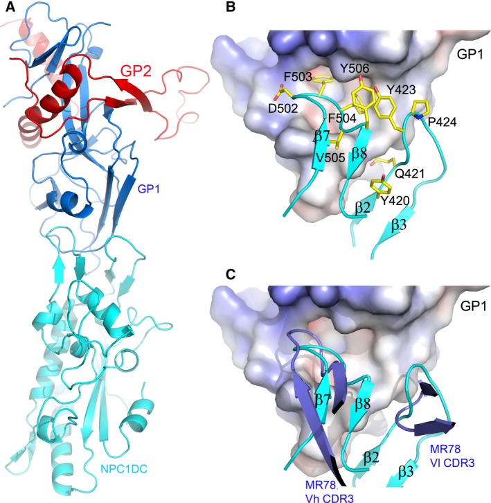
The docked complex of NPC1DC and EBOV GP. (A) Cartoon representation of NPC1DC (cyan) and EBOV GP (GP1, blue; GP2, red) complex. (B) Molecular interface of EBOV GP and NPC1DC. GP is shown as an electrostatic surface, NPC1DC as cyan ribbons with side‐chains as yellow sticks. (C) The docked NPC1DC and EBOV GP complex shows that β7–β8 and β2–β3 hairpins (cyan) are mimicked by interactions of the Vh CDR3 and Vl CDR3 (blue) of the antibody MR78, respectively, with Ebola GP.