
Figure 5.
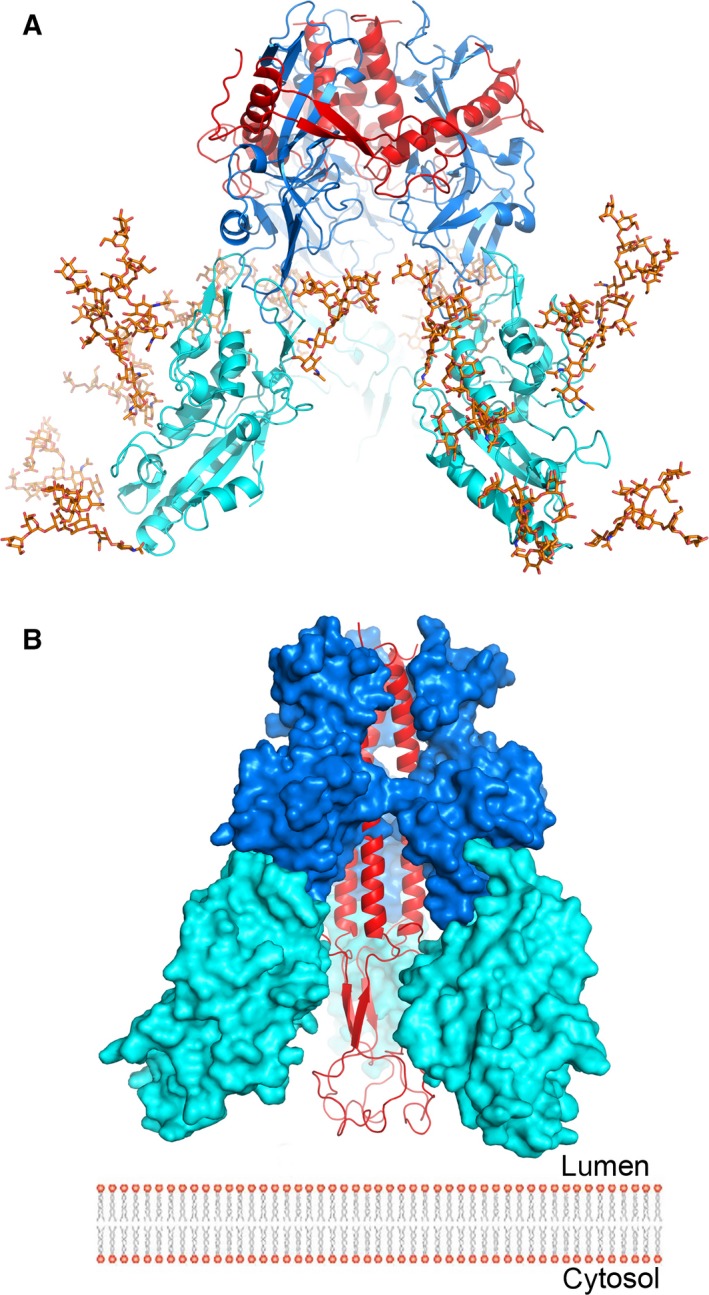
The putative trimeric complex of EBOV GP and NPC1DC. (A) The docked trimetric complex of Ebola GP and NPC1DC. GP1, GP2 and NPC1DC are coloured in blue, red and cyan, respectively. Man9GlcNAc2 is modelled at all glycosylation sites of NPC1DC showing that the glycans do not hinder the formation of the complex. (B) Subsequent to receptor binding, it is proposed that GP2 undergoes conformational changes. Its two helices (residues 553–597) coalesce to present the fusion loop to the endo/lysosomal membrane. The colour scheme is as in (A).