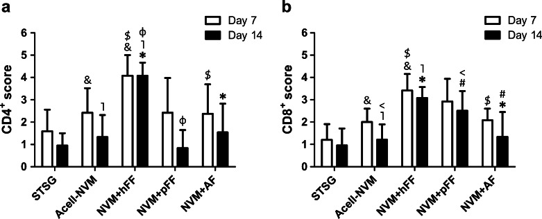
Fig. 5.
Influx of CD4+ and CD8+ lymphocytes in full-thickness wounds. The influx of CD4+ and CD8+ lymphocytes in the wounds was scored blinded for each treatment group (on a scale of 0 (none) to 5 (massive influx)). a Score of CD4+ lymphocyte influx in the wound area. A statistically significant higher influx of CD4+ lymphocyte was observed in wounds transplanted with NVM+hFF compared to Acell-NVM (&, ˥), and NVM+AF ($, *) (days 7 and 14). b Influx scores of CD8+ lymphocytes infiltrated into the wound area. Higher influx of CD8+ lymphocytes was observed in wounds treated with NVM+hFF or NVM+pFF in comparison to Acell-NVM (vs. hFF: &, ˥; vs. pFF: <) and NVM+AF (vs. hFF:$, *; vs. pFF: #). Statistical significance is indicated with symbols (Mann–Whitney U test, p < 0.05)

