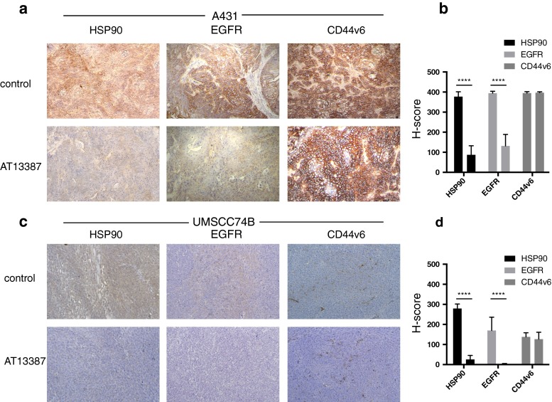
Fig. 5.
a c Ex vivo immunohistochemical staining for HSP90, EGFR and CD44v6 expression on representative sections of a A431 and c UM-SCC-74B tumour xenografts (×10). A431 tumours show high expression and UM-SCC-74B tumours show low expression of EGFR and CD44v6. HSP90 and EGFR were downregulated in the AT13387 treatment group. CD44v6 expression was unchanged. b, d Semiquantitative analysis of immunostaining using the H-score (n = 16, error bars SD; ****p < 0.0001, one-way ANOVA with Tukey’s post hoc test)

