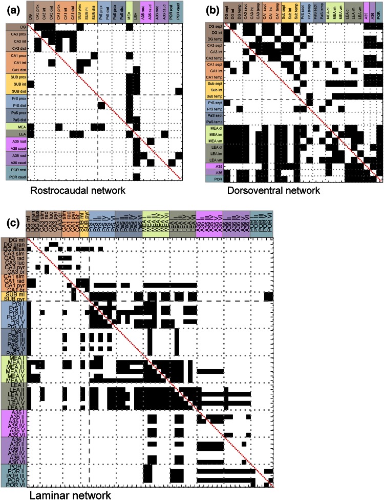
Fig. 1.
The directed, binary adjacency matrix of the rostrocaudal (a), dorsoventral (b) and laminar (c) network. The connections (or edges), presenting the axons or axon bundles, are indicated by black squares. The white areas represent projections that do not exist or are not documented yet. The self connections on the main diagonal (indicated with a red line) are excluded. The nodes represent the subareas of brain regions in the rostrocaudal dimension (a) or the dorsoventral dimension (b) and the layers of the brain regions (c). The nodes depicted on the left side represent the origin of a projection and the nodes on top represent the termination of a projection. The brain areas are color coded as follows: dentate gyrus (DG; dark brown), CA3 (medium brown), CA1 (orange) and subiculum (Sub; yellow), presubiculum (PrS; medium blue), parasubiculum (PaS; dark blue), entorhinal cortex (MEA; light green and LEA; dark green), perirhinal cortex [Brodmann areas (A) 35 (pink) and 36 (purple)] and the postrhinal cortex (POR; blue green). The Roman numerals indicate cortical layers. caud caudal, dist distal, dl dorsolateral part of the entorhinal cortex, gran granule cell layer, im intermediate dorsolateral–ventromedial part of the entorhinal cortex, luc stratum lucidum, ml molecular layer, or stratum oriens, prox proximal, pyr pyramidal cell layer, rad stratum radiatum, rost rostral, sept septal, slm stratum lacunosum-moleculare, temp temporal, vm ventromedial part of the entorhinal cortex

