
Figure 4. The proliferation and cytokine production of MART1- specific TILs in the presence of lovastatin.
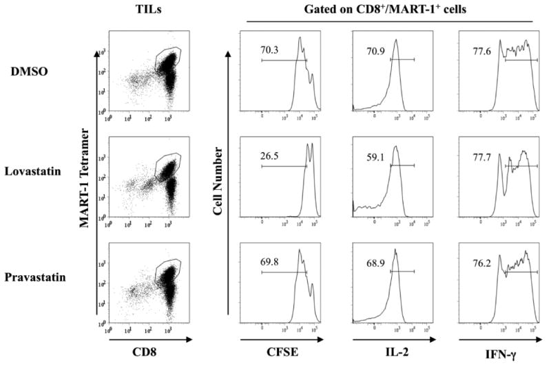
TILs from HLA-A2.1+ patients that recognize the HLA-A2.1-restricted MART-1 epitope were activated with mature dendritic cells pulsed with peptide in the presence of 10 μM lovastatin, 10 μM pravastatin or vehicle control DMSO as indicated. On day four of the stimulation, cells were collected and stained with surface markers to measure cell proliferation and cytokine production. MART-1 tetramer was used to identify antigen specific CD8+ T cells and CFSE was used to track the proliferation of gated cells. For IL-2 and IFN-γ production, TILs were coincubated with peptide–pulsed cells for 5 to 6 hours followed by a standard intracellular cytokine staining protocol. In each case, stained cells were analyzed in a FACScanto II flow cytometer (BD Biosciences). Isotype controls were used for setting the gates to determine the IL-2 and IFN-γ production. Data are representative of three individual TILs.