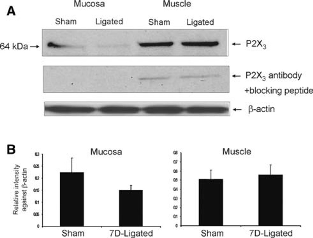
Fig. 5.
Western blot analysis of esophageal mucosa and muscle for P2X3 receptor protein expression in reflux esophagitis (7D-ligated) and sham-operated rats. β-actin immunostaining was used as a reference protein. a P2X3 was expressed as a 64 kDa protein both in esophageal mucosa and muscle extracts. Preabsorption of P2X3 antibody with immunogen (10 µM) completely eliminated the immunoreactivity in the mucosa and 90% in the muscle preparation. b Histograms of relative P2X3 band density in the experimental and control groups were normalized to β-actin staining. The results were expressed as mean ± SD of three experiments

