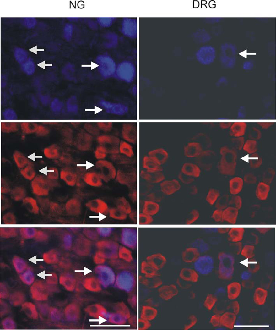
Fig. 7.
Photomicrography of P2X3 immunoreactivity in the NG and thoracic (T8) DRG retrogradely labeled with Fast Blue (FB). FB was injected in the subdiaphragmatic segment of the esophagus and FB positive cell bodies in the NG (left column) and DRG (right column) were examined for immunoreactivity with P2X3 antibody. Arrows indicate cells in NG and DRG with FB and P2X3 co-localization. The scale bar is 50 µm

