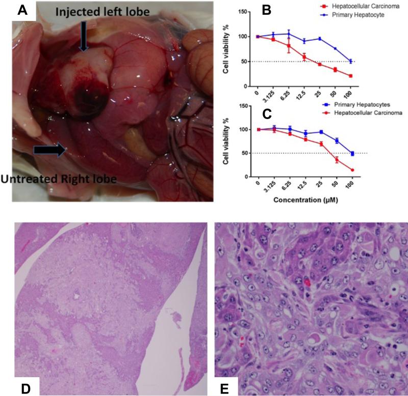
Figure 11.
(A) Left lobe of mouse liver injected with transformed hepatocytes indicating the development of tumor, whereas, the untreated right lobe appeared normal; In vitro assessment of drug activity as a free small molecule bexarotene (B) or PBNB (C) by using MTT assay at 72h; H&E stained sections of mouse liver transplanted with transformed hepatocytes. (D) Dissecting through the liver show sheets of transplanted cells. Bile ducts exhibit hyperplasia (1.2X); (E) transplanted cells arranged in sheets and trabeculae and supported by a thick fibrous stroma. Individual transplanted cells round to polygonal with moderate quantities of light eosinophilic homogenous cytoplasm and surrounded by indistinct cell border. Nuclei appeared single to multiple, round to oval with scattered chromatin and a single prominent central nucleolus. Small numbers of neutrophils look scattered within these regions. Existing trapped hepatocytes exhibit atrophy (3.40X).

