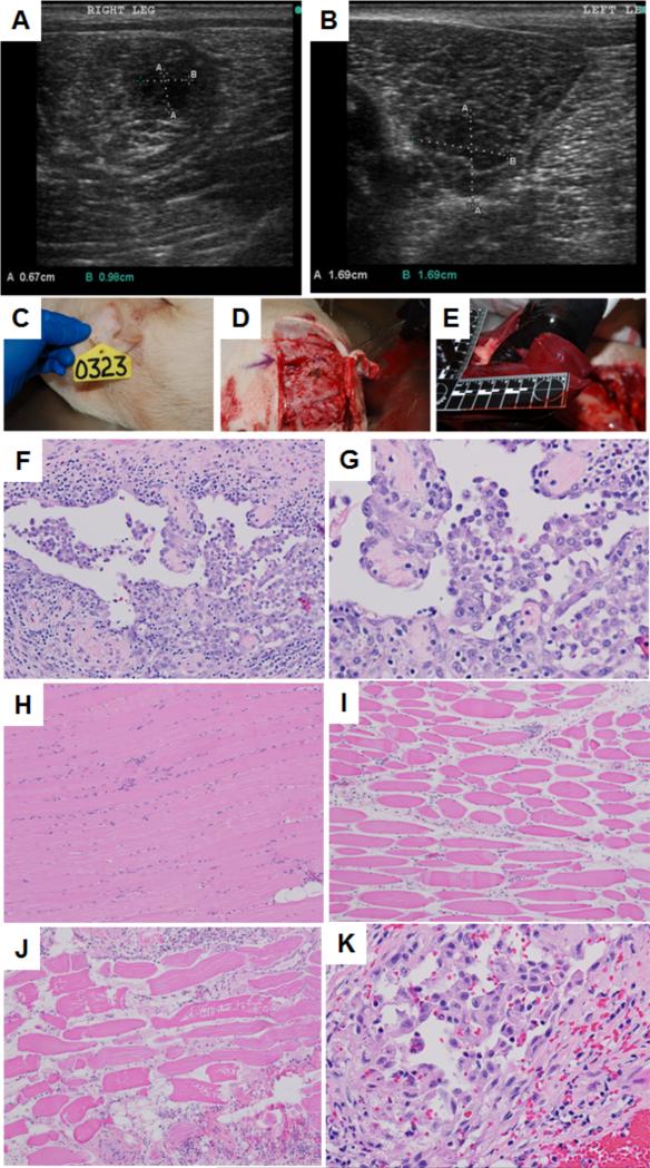
Figure 12.
Representative US images of abnormal tissue growth in (A) right and (B) left thigh of a transgenic pig. Representative necropsy images: (C) animal identification tag; (B) surgery at site of US exposure; (C) gross pathology of liver tissue; Histopathologic images: (F) neoplastic cells in the tumor from the left thigh showing lining vascular channels and caverns (20X), (G) neoplastic cells in the tumor from the right thigh showing lining vascular channels and caverns (40X), (H) skeletal muscle bundles not treated with thermal ablation (10X), (I) skeletal muscle fibers treated with thermal ablation exhibit sarcoplasmic hyalinization, loss of striation, cell shrinkage and increased endomysial space (10X), (J) skeletal muscle fibers treated with thermal ablation exhibit sarcoplasmic hyalinization, loss of striation, cell shrinkage, sarcoplasmic fragmentation and increased endomysial space (10X), (K) neoplastic cells in the tumor from the right thigh exhibit mild cytoplasmic swelling and vacuolation indicating vacuolar degeneration from the treatment (40X).

