
Fig. 1.
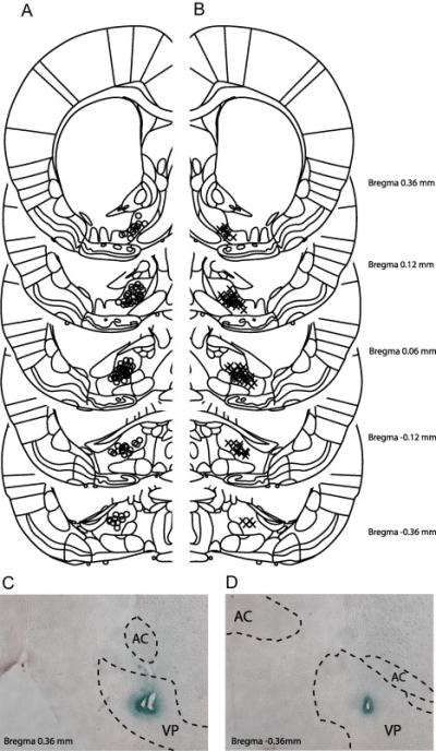
Electrophysiological recording sites in the VP. Histologically identified placements of electrode tips are depicted as Os for animals in the Cocaine group in (A), and Xs for animals in the Saline group in (B). Histological examples of electrode placements in the anterior (C) and posterior (D) VP are presented.