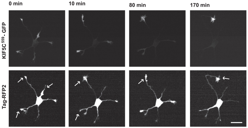
FIGURE 4. The Kinesin-1 motor domain translocates into different neurites until the axon is specified.
Selected frames from a time-lapse recording of a hippocampal neuron expressing constitutively active Kinesin-1 (KIF5C559-GFP) along with a soluble protein (Tag-RFP2) to visualize the cell’s geometry. Upper panels show the accumulation of the kinesin motor domain while lower panels illustrate the cell’s morphology; arrows denote neurites with significant kinesin accumulation. Prior to axon specification, the motor domain accumulates in two or three of the cell’s five neurites. As one neurite begins the period of extended growth that marks it as the axon, Kinesin-1 accumulates exclusively in this neurite, losing its ability to accumulate at the tips of any other neurites. Scale bar: 20 μm.

