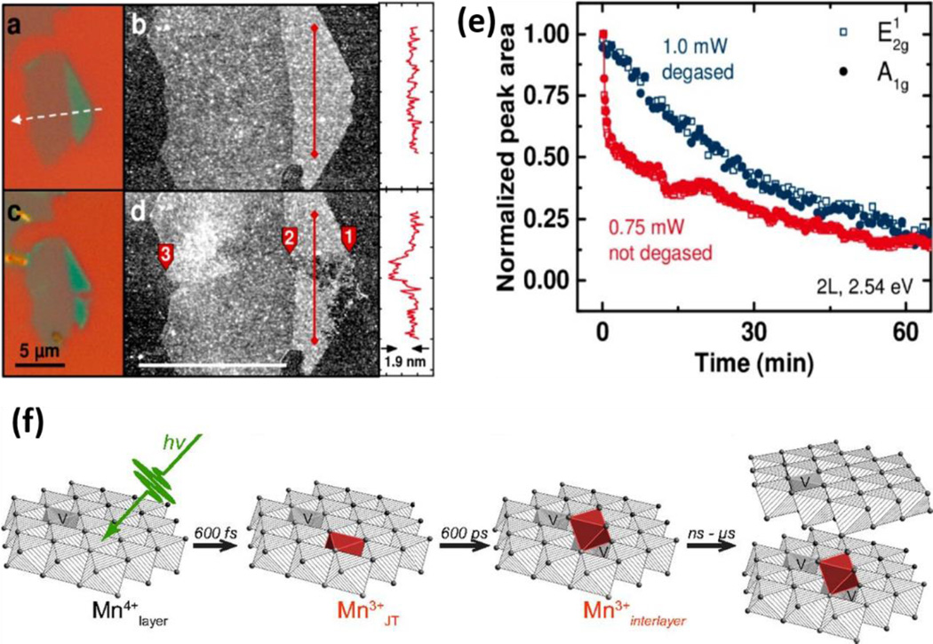
Figure 11.
(a, c) Optical microscopy (100×) and (b, d) AFM image of exfoliated MoS2 nanosheets before (top) and after (bottom) laser line scan, showing edge sites are the primary targets of photodegradation. e) Normalized Raman peak area as a function of illumination time on the edge site of a bilayer flake in the electrolyte with reduced oxygen (blue) and natural amount of dissolved oxygen (red). Reprinted with permission from ref. 214. Copyright 2015 American Chemical Society. (e) Proposed model for the evolution of redox chemistry in the photoreduction of MnO2 monolayer including photon absorption, formation of distorted Mn (III), migration of Mn(III) to an adsorption site and increased nanosheet stacking. Copyright (2015) National Academy of Sciences, USA.

