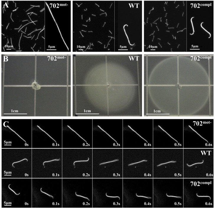
Fig 1. Morphology and motility of 702mot-, WT and 702compl strains.
(A) Observation under dark-field microscopy at ×20 or ×200 magnifications of cultures in liquid EMJH. (B) Spread of bacteria on soft 0.5% agar EMJH plates observed after 10 days of incubation. Identical results were obtained on 0.3% agar plates (S2 Fig). (C) Images taken every 0.1 s during 0.6 s under dark-field microscopy at ×200 magnification.

