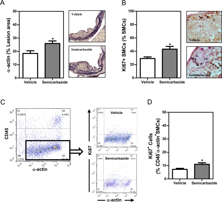
Fig 3. SSAO inactivation increased the percent of synthetic SMCs in established lesions under hypercholesterolemia.
Female LDLr KO mice were treated as described in the legend to Fig 1. (A) A bar graph (left) and representative photomicrographs (right) showing the lesion contents of SMCs. (B) A bar graph (left) or representative photomicrographs (right) showing the lesion contents of SMCs positive for Ki67. Sections of the aortic root were stained with the antibody against α-actin in the absence (A, purple for α-actin, 100x) or presence of anti-Ki67 (B, brown for α-actin while nuclear dark blue for Ki67, indicated by arrows, 400x) to visualize total or proliferative SMCs, respectively. Nuclei were counterstained by fast nuclear red (nuclear pink, B). (C) Dot plots showing proliferative VSMCs. Aortic cells were stained with antibodies against CD45, α-actin, and Ki67. The percent of proliferative (Ki67+) SMCs among α-actin+CD45- aortic cells are analyzed. (D) Bar graph showing the percent of proliferative (Ki67+) SMCs. Results were expressed as mean±SEM. Statistically significant difference *p<0.05 vs vehicle.

