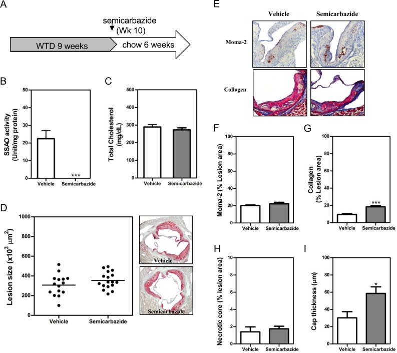
Fig 4. SSAO inactivation stabilized the established atherosclerotic lesions after normalization of hypercholesterolemia by diet lipid lowering.
Male LDLr KO mice were fed WTD for 9 weeks to induce the formation of atherosclerotic lesions. Thereafter, these animals were fed regular chow diet to normalize hypercholesterolemia (A). Drinking water containing 0.125% semicarbazide was given to these chow-fed animals in the subsequent 6 weeks (A) before the analysis of SSAO activities in the visceral adipose tissue (B), plasma cholesterol levels (C) or atherosclerotic lesions at the aortic root (D-G). (D) A scattered dot plot (left) or representative photomicrographs showing the size of atherosclerotic lesions. Sections were stained with oil-red-O (original magnification 40x, right panel). Each dot represents the mean lesion area of a single mouse, and the horizontal bar indicates the mean value of the group (left panel). (E) Representative photomicrographs showing the lesion contents of Moma-2+ macrophages (upper panel) or collagens (lower panel) in each group. Sections of aortic roots were stained with antibodies against Moma-2 to visualize macrophages (brown, 100x) or with Masson’s Trichrome Accustain to visualize collagens (blue, 100x). (F & G) Bar graphs showing the lesion contents of macrophages (F), collagens (G), necrotic core (H), and cap thickness (I) in vehicle- or semicarbazide-treated mice. Results were expressed as mean ±SEM. Statistically significant difference ***p<0.001 and *p<0.05 vs vehicle.

