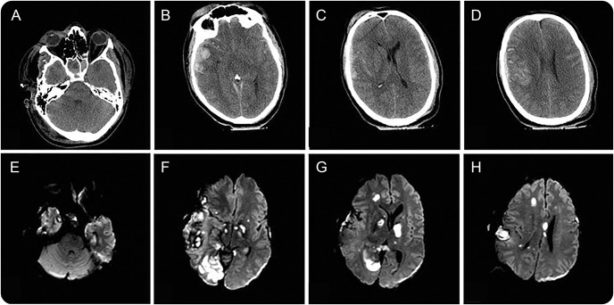
Figure 1. CT head and MRI brain.
Admission head CT shows right temporal contusion (B–C) with bilateral subarachnoid hemorrhage (B–D). Axial diffusion-weighted imaging 2 weeks later shows new infarcts in the right frontal lobe (G and H), bilateral deep nuclei and splenium of the corpus callosum (G), and right occipital lobe (F and G), while sparing the brainstem (E); prior right temporal contusion, subdural and subarachnoid hemorrhages, and areas of axonal injury are also noted.

