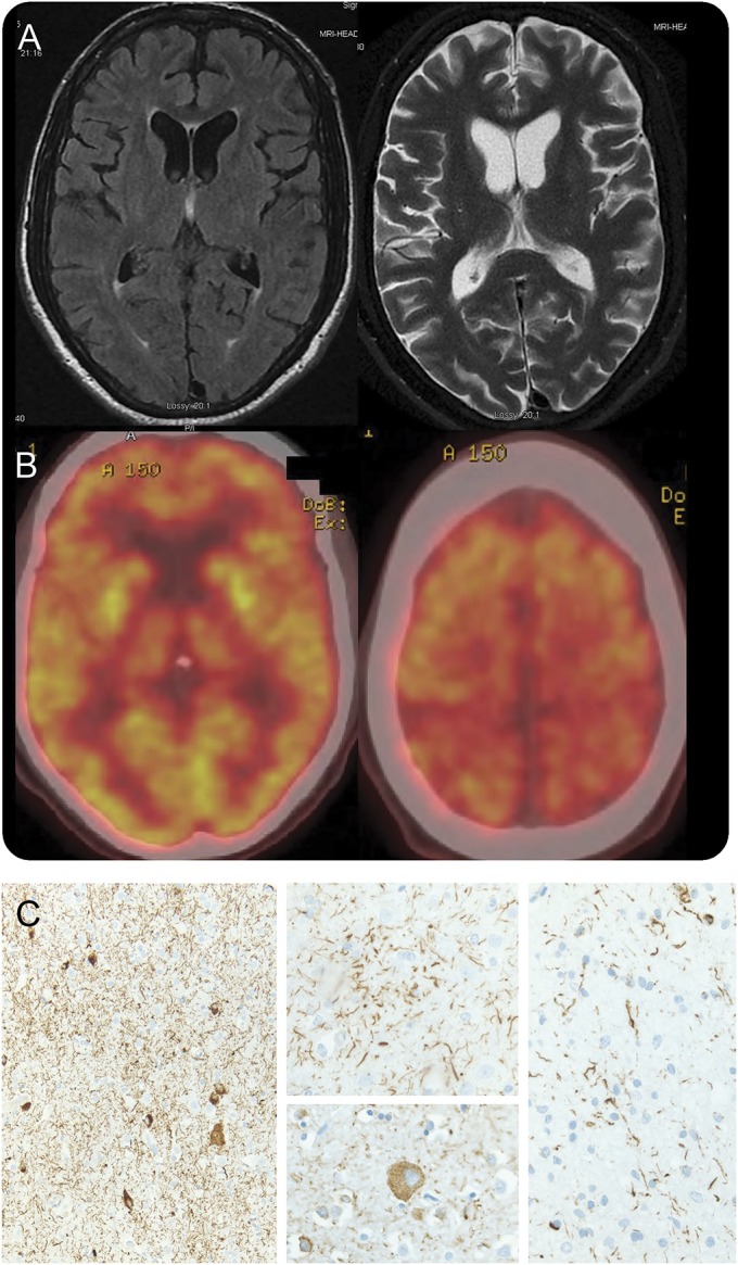
Figure. Selected brain MRI, fluorodeoxyglucose PET, and histopathology images.

(A) Axial brain MRI demonstrates mild atrophy of the left posterior frontal and anterotemporal lobes, widening the sylvian fissure. There was no abnormal signal in T1 or T2/fluid-attenuated inversion recovery sequences. (B) Fluorodeoxyglucose PET shows mild symmetric decreased metabolic activity in the posterior parietal lobes near the vertex, as well as in the thalami. (C) Histopathology (tau immunohistochemistry) demonstrated abnormal tau deposition within neurons and glia associated with prominent thread-like deposits within gray and white matter. Frontal cortical gray matter (left; original magnification 100×). Astrocytic plaque (center top) and balloon neuron (center bottom; original magnification 400×). White matter with coiled bodies and thread-like deposits (right; original magnification 400×).
