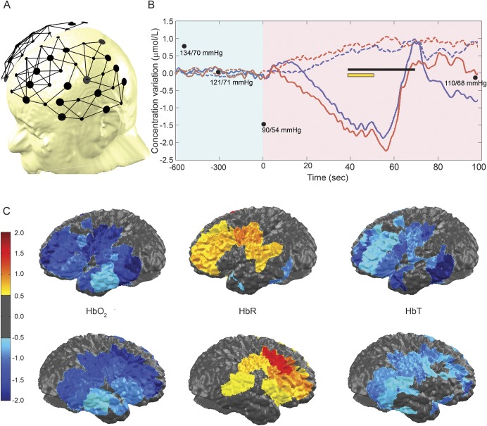
Figure 1. Functional near-infrared spectroscopy findings during the second episode of limb-shaking (LS) TIA.
(A) Channel configuration. (B) Hemodynamic variations over the dorsolateral frontal cortex and motor areas: no significant variations in total hemoglobin (HbT), oxyhemoglobin (HbO2), or deoxyhemoglobin (HbR) are found during a period of 10 minutes after standing up (condensed in light-shaded blue); progressive decrease in HbT and HbO2 and rise in HbR at t = 0 seconds (light-shaded purple). The black line indicates the period during which the patient experienced LS and lower limb weakness. The yellow line indicates the period during which we averaged hemodynamic changes over the frontal, temporal, and parietal cortices for topographic representation. Solid color line: HbO2. Red: right. Blue: left. Black dots: blood pressure. (C) Topographic views of HbO2, HbR, and HbT averaged from all ipsilateral channels between time 40 to 50 seconds.

