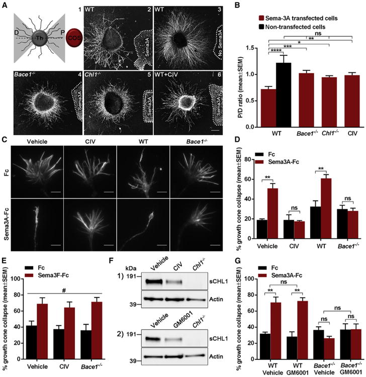
Figure 1. BACE1 Is Required for Sema3A-Induced Growth Cone Collapse in Co-culture Explants and Thalamic Neurons.

(A and B) Analysis of axonal sensitivity to Sema3A where WT (n = 17; A2), Bace1−/− (n = 18; A4), Chl1−/− (n = 15; A5), and BACE1-inhibitor-treated (1 mM CIV; n = 15; A6) thalamic explants were co-cultured with Sema3A-secreting aggregates of COS-1 cells. WT thalamic explants were also co-cultured with non-transfected COS-1 aggregates (n = 7; A3). The level of Sema3A-induced repulsion is represented by a P/D ratio, which compares axonal growth on proximal (P) and distal (D) side of the thalamic explant toward the COS aggregate (A1).
(C and D) Analysis of Sema3A-induced growth cone collapse in WT, BACE1-inhibitor-treated (1 μM CIV), and Bace1−/− thalamic neurons.
(E) Quantification of Sema3F-induced growth cone collapse in WT, BACE1-inhibitor-treated (1 μM CIV), and Bace1−/− thalamic neurons.
(F) Western blot analysis of WT thalamic neurons treated with 1 μM CIV (1) or with 50 μM GM6001 (2).
(G) Quantification of Sema3A-induced growth cone collapse in WT and Bace1−/ − thalamic neurons treated with 50 μM GM6001.
Results are presented as mean ± SEM. The scale bars represent 200 μm (explants) and 5 μm (growth cones). See also Figure S1.
