
Fig 6.
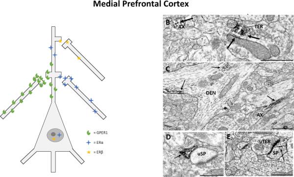
A) Representation of estrogen receptor localization to pre- and post-synaptic profiles in the prefrontal cortex. Estrogen receptors were most commonly localized to axons and terminals in the prefronal cortex, depicted alongside the pyramidal neuron, and were also observed in dendrites and dendritic spines at lower levels, depicted on the apical dendrite of the pyramidal neuron. Low levels of nuclear labelling for ERα and ERβ were observed via light microscopy. B) ERα in an axon (AX) and in a terminal (TER), where immunoreactivity (IR) is observed at small synaptic vesicles and on the membrane of a mitochondrion (mit), C) GPER1-IR in an axon (AX) and in a dendrite (DEN),where it is associated with microtubules and the cell membrane, D) ERβ-IR in a glial cell (GL) that is in apposition to an unlabeled dendritic spine (uSP), E) ERβ-IR in a dendritic spine (SP) that forms an asymmetrical synapse with an unlabeled terminal (uTER). Black arrow, immunoperoxidase labeling for estrogen receptors; Scale Bar, 500nm. Taken from Almey, A., Cannell, E., Bertram, K., Filardo, E., Milner, T.A., Brake, W.G.. 2014. Medial prefrontal cortical estradiol rapidly alters memory system bias in female rats: ultrastructural analysis reveals membrane-associated estrogen receptors as potential mediators. Endocrinology 155, 4422-32.