
Fig. 1.
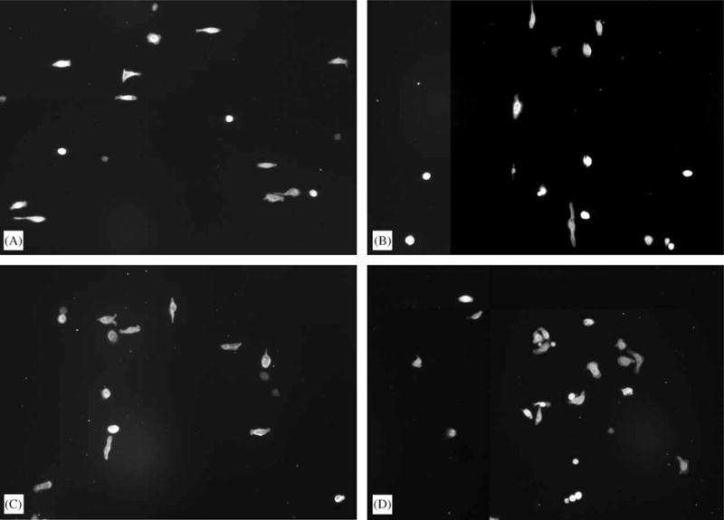
Fluorescence images of cells stained for actin. The patterns were oriented in the vertical direction. (A) On 70 nm ridges on a 400 nm pitch, cells aligned preferentially in the direction perpendicular to the patterns. (B) Cells aligned in the direction of the patterns on 1900 nm ridges on a 4000 nm pitch. (C) On 400 nm ridges on a 1200 nm pitch, cells aligned both parallel and perpendicularly to the patterns. (D) On smooth substrates cells were randomly oriented.