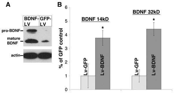
Fig. 2.
BDNF-lentivirus increases BDNF levels within the spinal cord. Lentivirus encoding for BDNF or GFP was injected into normal C7 spinal cord tissue. (A) Western blot analysis indicates that three weeks after the injection, BDNF levels (~14 kD) were approximately 3.8 times higher in tissue in the animals that were injected with BDNF-lentivirus (dark bars in B) than GFP-lentivirus (light bars in B). Moreover, there was also an increase (~4.4×) in the expression of the immature, precursor BDNF (~32 kD) in these animals. Y-axis values in B are the ratio of the densitometric values (following normalization to actin) to those of the GFP-lentivirus control (mean±SEM). *p<0.05 between BDNF-lentivirus and GFP-lentivirus.

