
Fig. 3. Effects of genetic deletion of TLR9 on adipose tissue inflammation and insulin resistance.
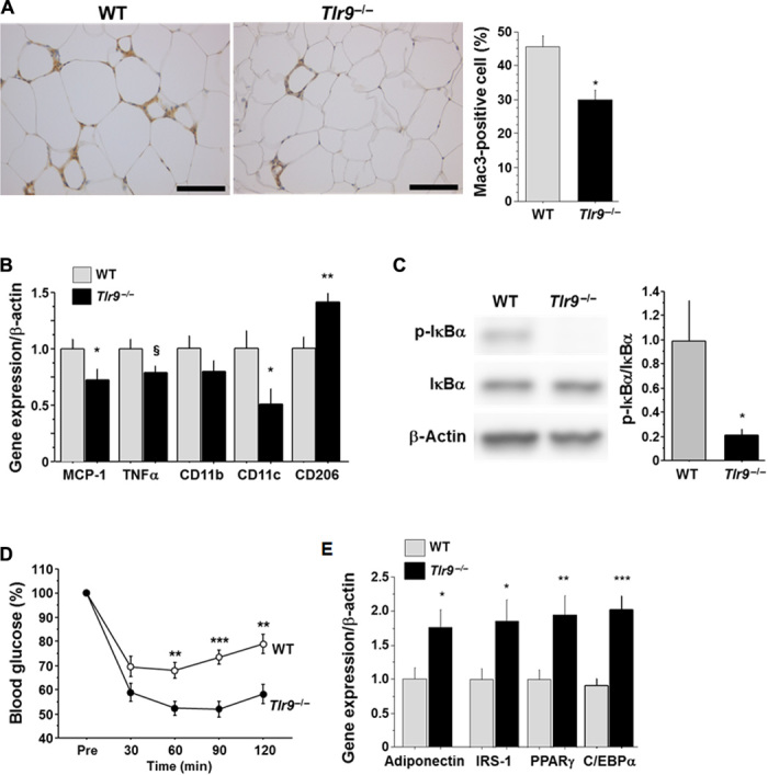
(A) Mac3 staining of epididymal fat obtained from WT or Tlr9−/− mice (n = 12 to 15). Scale bar, 100 μm. (B) Quantitative RT-PCR analysis of inflammatory gene expression in epididymal fat obtained from WT or Tlr9−/− mice (n = 12). (C) Western blot analysis of the phosphorylation of IκBα in epididymal fat obtained from WT or Tlr9−/− mice (n = 11). (D) Results of the insulin tolerance test (0.75 U/kg) of WT or Tlr9−/− mice (n = 13 to 16). (E) Quantitative RT-PCR analysis of epididymal fat for genes related to insulin sensitivity (n = 12). C/EBPα, CCAAT/enhancer binding protein α. All experiments in this figure were performed using samples obtained from mice fed a high-fat diet (HFD) for 12 weeks. §P = 0.05, *P < 0.05, **P < 0.01, and ***P < 0.001. All values are means ± SEM.