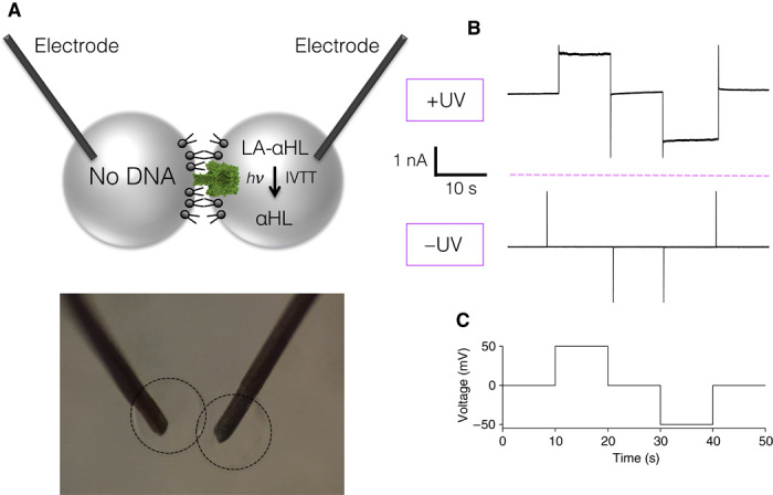
Fig. 4. Light-activated electrical signal between synthetic cells.

(A) Schematic of the synthetic cell pair. One cell contains LA-αHL DNA, the other contains no DNA. Both droplets have electrodes inserted within them to apply a potential and measure the ionic current. Below is an image of the experimental setup. (B) A current is detected only following the expression of αHL after light activation. (C) Voltage protocol used in (B).
